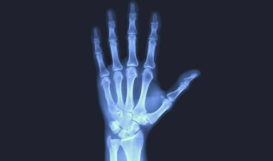
Radiography equipment consists of a large, flat table with a drawer that holds a tray into which an x-ray film cassette is placed.

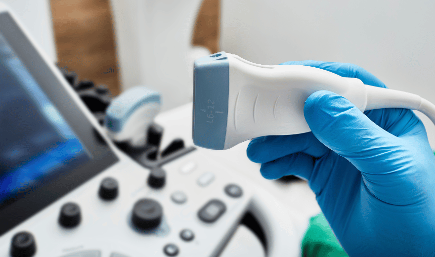
involves sending sound waves into the body. These sound waves reflect off the internal organs and are recorded by special instruments that create images of anatomic parts.
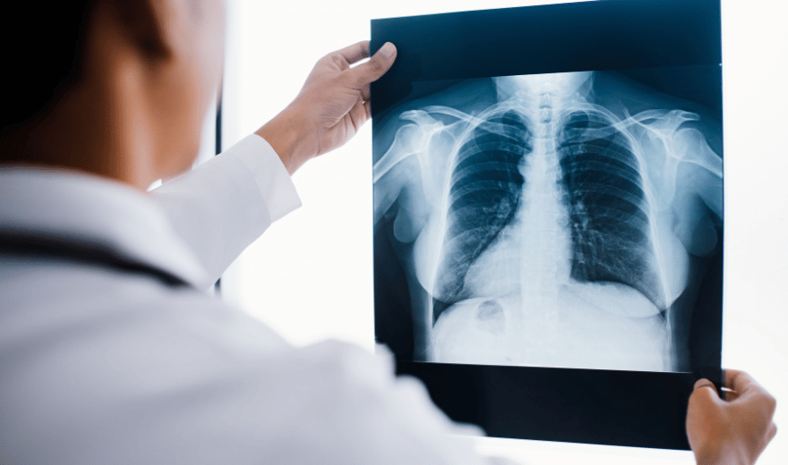
A radiologist (a physician experienced in ultrasound and other radiology examinations) will analyze the images and send a signed report with his or her interpretation to the patient’s personal physician.
This is a painless procedure. The primary discomfort may come from the coldness of the recording plate.

Do not wear deodorant, talcum powder, or lotion under your arms or on your breasts on the day of the exam. These can appear on the x-ray film as calcium spots.
pelvic ultrasound is most often used to examine the uterus and ovaries and, during pregnancy, to monitor the health and development of the embryo or fetus
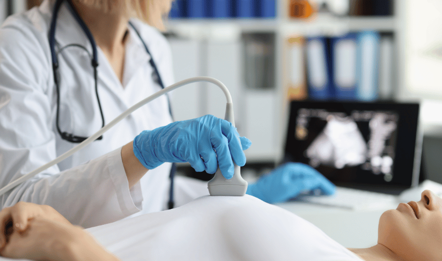
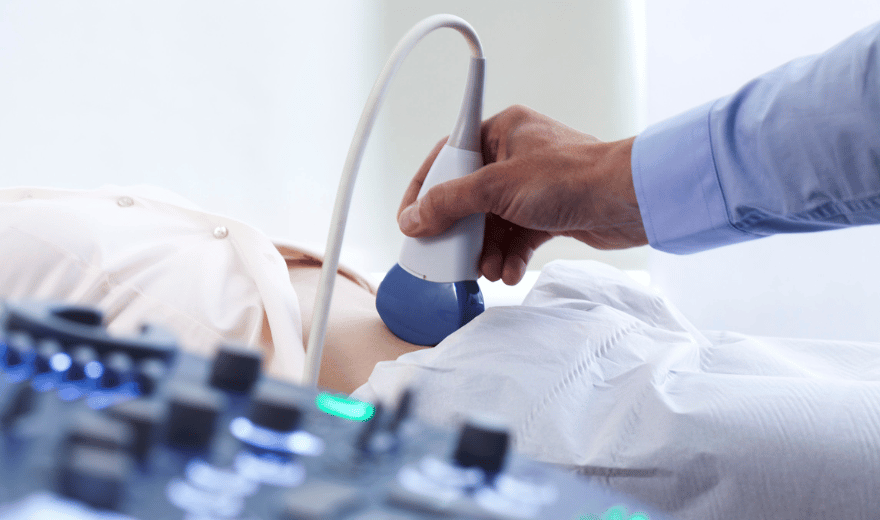
Ultrasound imaging, also called ultrasound scanning or sonography, involves exposing part of the body to high-frequency sound waves to produce pictures of the inside of the body.
Ultrasound or sonography involves the sending of sound waves through the body. Those sound waves are reflected off the internal organs.
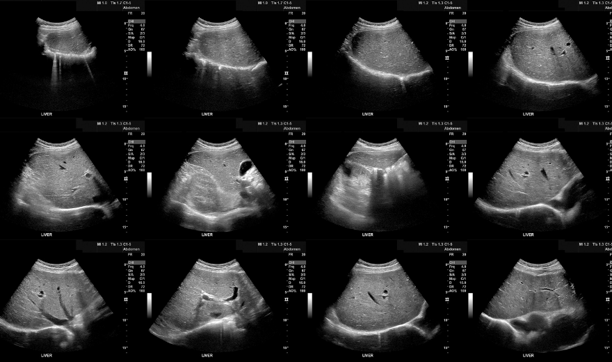
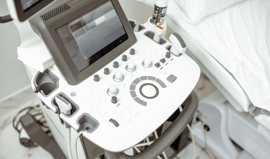
An abdominal ultrasound image is a useful way of examining internal organs, including the liver, gallbladder, spleen, pancreas, kidneys, and bladder.
If an ultrasound is required early in your pregnancy, you will be required to have a full bladder for the procedure.
Services
Site Links
© Copyright 2012 - 2026 • Annex Medical Imaging • Designed and Developed by White Dolphin Digital • All Rights Reserved