WHAT IS ULTRASOUND IMAGING OF MUSCULOSKELETAL SYSTEM?
Musculoskeletal ultrasound or sonography involves sending sound waves into the body. These sound waves are reflected off the internal organs and are then interpreted by special instruments that create an image of anatomic parts. No ionizing radiation (x-ray) is involved in ultrasound imaging. Ultrasound images are captured in real-time so they can show movement of internal tissues and organs and the flow of blood in the arteries and veins.
An ultrasound image is a useful way of examining the musculoskeletal system of the body to detect problems with muscles, tendons, ligaments, joints and soft tissue. Ultrasound images are captured in real time, so they can often show movement, function and anatomy, as well as enable radiologists to diagnose a variety of conditions and assess damage after an injury or illness.
WHAT ARE SOME COMMON USES OF THE PROCEDURE?
Ultrasound images can be useful in diagnosing tendon tears, such as tears of the rotator cuff in the shoulder or Achilles tendon in the ankle. Abnormalities of the muscles can also be seen, such as tears and soft-tissue masses. Bleeding or other fluid collections within the muscles, bursae and joints can also be detected. Ultrasound has not proven useful in the diction of whiplash injuries or other causes of back pain.
HOW SHOULD I PREPARE FOR THE PROCEDURE?
You should wear comfortable, loose-fitting clothing for your ultrasound exam. No other preparation is required. You will have to remove clothing and jewellery in the area to be examined.
HOW DOES THE PROCEDURE WORK?
The ultrasound transducer generates sound waves that pass through the skin and also serves as a microphone to record the returning sounds- the echoes. When pressed against the skin, the transducer directs high frequency sound waves toward the structures being studied and records any changes in the pitch and direction of the returning echoes. The bounce-back echoes, or the signature are automatically measured by the computer and converted electronically to a picture that shows what is happening at that instant- creating a so called- ‘real-time’ image on the monitor screen. These images are frozen in time to obtain still pictures. If a Doppler study is done, blood flow can be displayed in color on the screen and actually heard.
WHAT WILL I EXPERIENCE DURING THE PROCEDURE?
Most ultrasound studies are relatively quick and well-tolerated by the patient. If scanning is performed over an area of tenderness, there may be minor pain associated with the procedure. Otherwise the procedure is painless.
WHAT ARE THE BENEFITS VS. RISKS?
Benefits
-
Ultrasound scanning is usually painless and non-invasive.
-
Ultrasound is widely available and easy to use.
-
Ultrasound imaging uses no ionizing radiation.
-
Ultrasound provides real-time imaging, making it a good tool for guiding minimally invasive procedures, such as cortisone injections, needle biopsies and aspiration of fluid in joints or elsewhere.
-
Unlike the strong magnetic field of magnetic resonance imaging (MRI), ultrasound is not affected by cardiac pacemakers, ferromagnetic implants or fragments within the body. Ultrasound is also an excellent alternative to MRI for claustrophobic patients.
-
Ultrasound may actually have advantages over MRI in seeing tendon structure, which is better appreciated by ultrasound than MRI.
Risks
For standard diagnostic ultrasound there are no known harmful effects on humans.
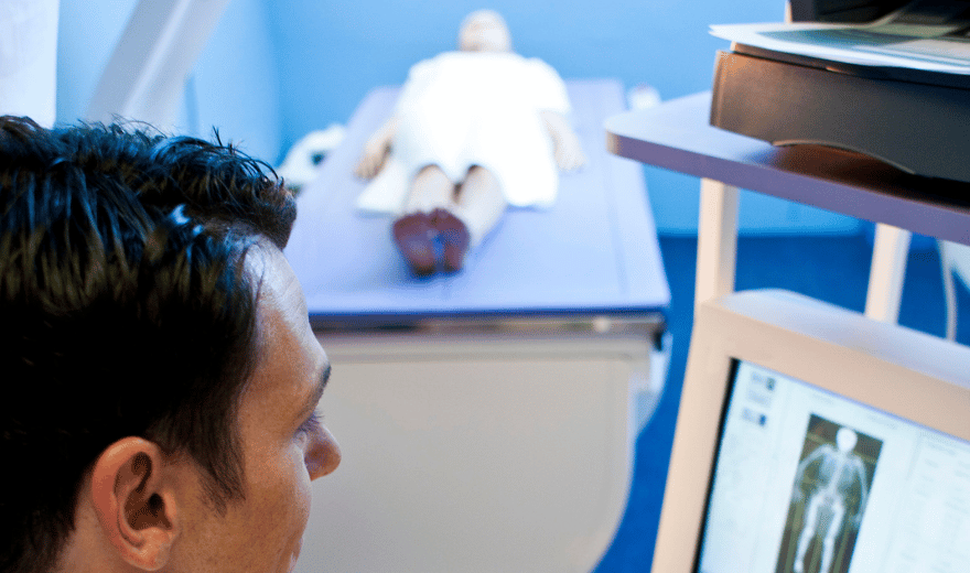
WHAT DOES THE EQUIPMENT LOOK LIKE?
The equipment consists of a transducer and a monitoring system. The transducer is a small, hand-held device that resembles a microphone. The radiologist or sonographer spreads a lubricating gel on the area being examined and then presses this device firmly against the skin.
The ultrasound image is immediately visible on a nearby screen that looks much like a computer or television monitor. The radiologist or sonographer watches this screen during an examination and captures representative images for storage. Often the patient is able to see it as well.
HOW IS THE PROCEDURE PERFORMED?
The patient is positioned on an examination table. A clear gel is applied to the area that will be examined. The gel helps the transducer make a secure contact and eliminates air pockets between the transducer and the skin since the sound waves cannot penetrate air. The sonographer then presses the transducer firmly against the skin and sweeps it back and forth to image the area of interest, reviewing the images on the monitor and capturing “snapshots” as required. The entire area of interest will be scanned obtaining images from different perspectives. Sometimes the examiner may want to obtain images while you are sitting or standing upright. The examination usually takes less than 45 minutes.
When the examination is complete, the patient may be asked to dress and wait while the ultrasound images are reviewed. Often, though, the sonographer or radiologist is able to review the ultrasound images in real time as they are acquired, and the patient can be released immediately.
Also Read: Pediatric abdominal ultrasound and preparation
WHO INTERPRETS THE RESULTS AND HOW DO I GET THEM?
A radiologist (a physician experienced in ultrasound and other radiology examinations) will analyze the images and send a signed report with his or her interpretation to the patient’s personal physician. The patient receives ultrasound results from the referring physician who ordered the test results.
WHAT ARE THE LIMITATIONS OF ULTRASOUND IMAGING OF THE MUSCULOSKELETAL SYSTEM?
Ultrasound has difficulty penetrating bone and, therefore, can only see the outer surface of bony structures and not what lies within and beyond. For visualizing internal structure of bones or certain joints, other imaging modalities, such as MRI, should be selected.
MUSCULOSKELETAL ULTRASOUND
Ultrasound or sonography involves sending sound waves into the body. These sound waves are reflected off the internal organs and are then interpreted by special instruments that create an image of anatomic parts. No ionizing radiation (x-ray) is involved in ultrasound imaging. Ultrasound images are captured in real-time so they can show movement of internal tissues and organs and the flow of blood in the arteries and veins.
An ultrasound image is a useful way of examining the musculoskeletal system of the body to detect problems with muscles, tendons, ligaments, joints and soft tissue. Ultrasound images are captured in real time, so they can often show movement, function and anatomy, as well as enable radiologists to diagnose a variety of conditions and assess damage after an injury or illness.
MUSCULOSKELETAL ULTRASOUND PREPARATION
These instructions are IMPORTANT. Please follow them.
-
You should wear comfortable, loose-fitting clothing for your ultrasound exam.
-
You will have to remove clothing and jewellery in the area to be examined.
-
Please let us know before your exam begins if you may be pregnant.
continue reading
Related Posts

Radiography equipment consists of a large, flat table with a drawer that holds a tray into which an x-ray film cassette is placed.

involves sending sound waves into the body. These sound waves reflect off the internal organs and are recorded by special instruments that create images of anatomic parts.