WHAT IS BONE RADIOGRAPHY?
Radiography, or an Bone X-Ray, as it is most commonly known, is the oldest and most frequently used form of medical imaging. Discovered more than a century ago, x-rays can produce diagnostic images of the human body on film or digitally on a computer screen.
X-ray imaging is the fastest and easiest way for a physician to view and assess broken bones. At least two images (from different angles) are taken and often three images are needed if the problem is around a joint (knee, elbow or wrist). X-rays also play a key role in guiding orthopedic surgery and in the treatment of sports-related injuries. X-ray may uncover more advanced forms of cancer in bones, although early screening for cancer findings requires other methods.
WHAT ARE SOME COMMON USES OF THE PROCEDURE?
Probably the most common use of bone radiographs is to assist the physician in identifying and treating fractures. Images of the injury can show even very fine hairline fractures or bone chips, while images produced after treatment ensure that a fracture has been properly aligned and stabilized for healing. Bone x-rays are essential tools in orthopedic surgery, such as spinal repair, joint replacements or fracture reductions.
X-ray images can be used to diagnose and monitor the progression of degenerative diseases such as arthritis. They also play an important role in the detection and diagnosis of cancer, although usually computed tomography (CT) or MRI is better at defining the extent and the nature of a suspected cancer. On regular x-rays severe osteoporosis can be visible, but bone density determination for early loss of bone mineral is usually done on specialized, more sensitive equipment.
Preparation
HOW SHOULD I PREPARE FOR THE PROCEDURE?
There is no special preparation required for most bone radiographs. Once you arrive, you may be asked to change into a gown before your examination. You will also be asked to remove jewelry, eyeglasses and any metal objects that could show up on the images and overlap important findings. Women should always inform their doctor, radiologist, and x-ray technologist if there is any possibility that they are pregnant.
HOW DOES THE PROCEDURE WORK?
Radiography involves exposing a part of the body to a small dose of radiation to produce an image of the internal organs. When x-rays penetrate the body, they are absorbed in varying amounts by different tissues. Ribs, for example, are dense and will block much of the radiation and, therefore, appear white or light gray on the image. Soft tissue such as the liver or lungs will appear darker because more radiation can pass through it to expose the film.
WHAT WILL I EXPERIENCE DURING THE X-RAY PROCEDURE?
X-ray imaging itself is painless. Some discomfort may result from lying on the table, a hard surface that may feel quite cold. Sometimes, to get a clear image of an injury such as a possible fracture, you may be asked to hold an uncomfortable position for a short time. Any movement could blur the image and make it necessary to repeat the procedure to get a useful, clear picture.
WHAT ARE THE BENEFITS VS. RISKS?
Benefits
- X-ray imaging is useful to diagnose bone injury and disease, such as fractures, bone infections, arthritis and cancer.
- Because x-ray imaging is fast and easy, it is particularly useful in emergency diagnosis and treatment.
- X-ray equipment is relatively inexpensive and widely available in physician offices, ambulatory care centers, nursing homes and other locations, making it convenient for both patients and physicians.
Risks
- X-rays are a type of invisible electromagnetic radiation and create no sensation when they pass through the body. Modern x-ray techniques use only a fraction of the x-ray dose that was required in the early days of radiology.
- Women should always inform their doctor or x-ray technologist if there is any possibility that they are pregnant.
- During a single x-ray exposure, a patient is exposed to approximately 20 milliroentgens of radiation. To put this into perspective, we are all exposed to approximately 100 milliroentgens of radiation each year from sources like the ultraviolet rays of the sun and small traces of radioactive isotopes, such as uranium found in soil.
Radiation risks are further minimized by:
- The use of high-speed x-ray film that requires only very small amounts of radiation to produce an optimal image.
- Technique standards established by national and international guidelines that have been designed and are continually reviewed by national and international radiology protection councils.
- Modern, state-of-the-art x-ray systems (including mammography systems, angiographic equipment, labs and CT scanners) that have very tightly controlled x-ray beams with significant filtration and x-ray dose control methods. Scatter or stray radiation is minimized and those parts of a patient’s body not being imaged receive minimal exposure
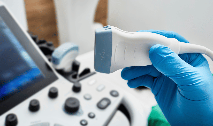
Also Read: Pediatric abdominal ultrasound and preparation
WHAT DOES THE X-RAY EQUIPMENT LOOK LIKE?
Radiography equipment consists of a large, flat table with a drawer that holds a tray into which an x-ray film cassette is placed. Suspended above the table is the apparatus that holds the x-ray tube that can be moved over the body to direct the x-ray.
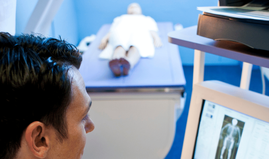
HOW IS THE PROCEDURE PERFORMED?
The technologist positions the patient on the examination table, places a film holder (cassette) under the table in the area of the body to be imaged. Sandbags or pillows may help the patient hold the proper position. Then the technologist steps behind a radiation barrier and asks the patient to hold very still without breathing for a few seconds. The radiographic equipment is activated, sending a beam of x-rays through the body to expose the film. The technologist then repositions the patient for another view, and the process is repeated.
When your x-rays are completed you will be asked to wait until the technologist checks the images for adequate exposure and motion.
WHO INTERPRETS THE RESULTS AND HOW DO I GET THEM?
A radiologist (a physician experienced in bone x-ray and all other types of radiology examinations) will analyze the images and send a signed report to your primary care or referring physician, who will inform you of your test results.
WHAT ARE THE LIMITATIONS OF BONE X-RAY RADIOGRAPHY?
While x-ray images are among the clearest, most detailed views of bone, they provide little information about the adjacent soft tissues. In the case of a knee or shoulder injury, for example, an MRI or ultrasound may be more useful in identifying ligament tears, joint effusions or other problems.
Even in the evaluation of a traumatic injury to the bone that does not cause a visible crack, MRI can detect a so-called bone “bruise.” Other imaging modalities, such as positron emission tomography (PET), bone scanning, or CT, may be more effective in diagnosing cancer metastases (spread) to bone or primary bone tumors. MR is especially useful for imaging the spine because the bones and the spinal cord are evaluated. Ultrasound (sound waves instead of ionizing radiation) has also been useful in injuries around joints and in evaluating hips in children for congenital problems.
continue reading
Related Posts

involves sending sound waves into the body. These sound waves reflect off the internal organs and are recorded by special instruments that create images of anatomic parts.
A radiologist (a physician experienced in ultrasound and other radiology examinations) will analyze the images and send a signed report with his or her interpretation to the patient’s personal physician.