WHAT IS CHEST RADIOGRAPHY?
Chest x-ray is the most commonly performed diagnostic x-ray examination. Approximately half of all x-rays obtained in medical institutions are chest x-rays. A chest x-ray is usually done for the evaluation of lungs, heart and chest wall. Pneumonia, heart failure, emphysema, lung cancer and other medical conditions can be diagnosed or suspected on a chest x-ray. Traditionally, chest x-rays have been taken prior to employment, prior to surgery or during immigration.
WHAT ARE SOME COMMON USES OF THE PROCEDURE?
Chest x-ray is typically performed as the first imaging test for symptoms of shortness of breath, a bad or persistent cough, chest pain, chest injury or fever. Individuals with known or suspected medical conditions such as congestive heart failure or cancer may have chest x-rays to follow their response to treatment, or to determine changes that would require a change in their medical management.
HOW SHOULD I PREPARE FOR THE PROCEDURE?
This procedure requires no special preparation. Women should always inform their doctor, radiologist, and x-ray technologist if there is any possibility that they are pregnant.
HOW DOES THE PROCEDURE WORK?
Radiography involves exposing a part of the body to a small dose of radiation to produce an image of the internal organs. When x-rays penetrate the body, they are absorbed in varying amounts by different parts of the anatomy. The ribs and spine, for example, absorb much of the radiation and appear white or light gray on the image.
Lung tissue absorbs little radiation and appears dark on the image. Chest x-rays can be maintained as hard copy film for filing or at Annex Medical Imaging as filmless digital images that are archived electronically. Digital images can also be transferred for storage onto CD-ROM. Stored images may be used to compare with later images if illness develops. Indeed, historical comparison films are often very important in the decision process as to whether a finding is clinically important or not.
WHAT WILL I EXPERIENCE DURING THE X-RAY PROCEDURE?
This is a painless procedure. The primary discomfort may come from the coldness of the recording plate. Individuals with arthritis or injuries to the chest wall, shoulders or arms may have discomfort trying to maintain position for the chest x-ray. In these circumstances, the technologist will assist you in finding a position that still ensures diagnostic image quality.
WHAT ARE THE BENEFITS VS. RISKS?
Benefits
- A physician may recommend a chest x-ray for a patient with shortness of breath, a bad or persistent cough, chest pain or a chest injury. In the instances of pneumonia, the site of pneumonia and fluid in the lungs will appear white on the image.
- A chest x-ray may also show advanced emphysema as well as other diffuse lung conditions, such as pulmonary fibrosis.
- Lung cancers and tumours that spread to the lung may be visible on chest x-ray. However, lesions that are very small or superimposed on normal structures may not always be visible.
- Heart enlargements caused by fluid around the heart (pericardial effusion), heart muscle disease, abnormal heart anatomy or congestive heart failure may also be visible on an x-ray depending on their severity.
- Pleural effusions (fluid around the lungs) on one or both sides can be detected. Usually the cause of such fluid may be deduced from clinical data or other findings on the chest x-ray; however, it may be necessary to sample the fluid to determine its nature.
Risks
- X-rays are a type of electromagnetic radiation, are invisible and create no sensation when they pass through the body. The chest x-ray is one of the lowest radiation exposure medical examinations performed today.
- Special care is taken during x-ray examinations to ensure maximum safety for the patient by paying attention to correct x-ray beam energies. Shielding the abdomen and pelvis with a lead apron helps reduce unnecessary radiation to the abdomen and pelvis. Women should always inform their doctor or x-ray technologist if there is any possibility that they are pregnant.
- The effective radiation dose from this procedure is about 0.1 mSv, which is about the same as the average person receives from background radiation in 10 days.
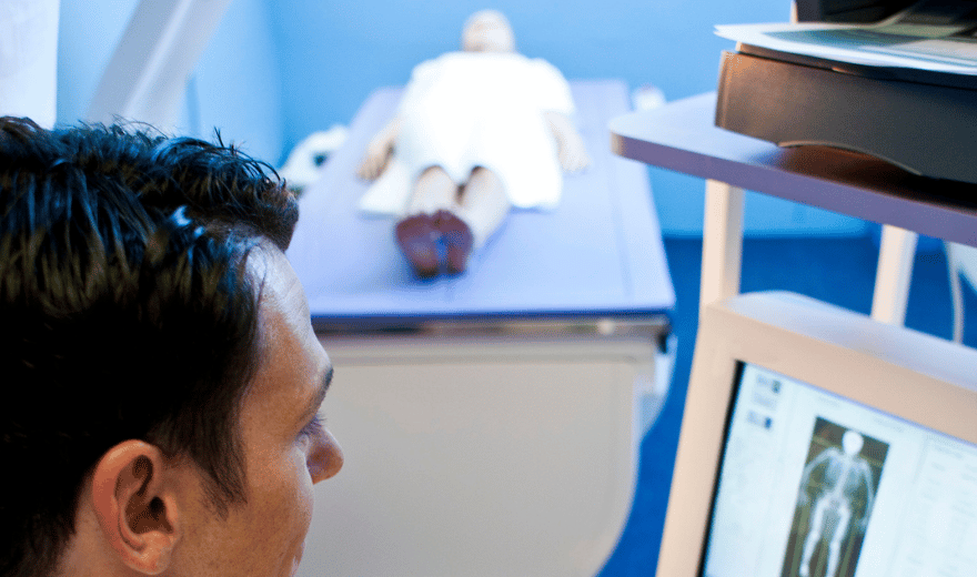
WHAT DOES THE X-RAY EQUIPMENT LOOK LIKE?
The most common radiography equipment used for chest x-rays consists of a box-like apparatus containing the recording material against which the individual places his/her chest-and the apparatus containing the x-ray tube, usually positioned about six feet away.
HOW IS THE PROCEDURE PERFORMED?
Patients must remove their clothing, including undergarments that may contain metal. You will be given a loose-fitting gown to wear. Patients will also be asked to remove all metallic jewelry that may interfere with the x-rays. Normally, a frontal or posteroanterior view is obtained, in which the patient stands with the chest pressed to the photographic plate, with hands on hips and elbows pushed in front in a somewhat exaggerated position.
The technologist will ask the patient to be still and to take a deep breath and hold it. Breath-holding after a deep breath reduces the possibility of a blurred image and also enhances the quality of the x-ray image, since abnormalities in air-filled lungs are easier to see than in deflated lungs.
Next, the technologist walks into a cubicle or small room to activate the radiographic equipment, which sends a beam of x-rays from the x-ray source behind the patient, through the patient’s chest, to the recording medium.
The technologist may need to take additional views to properly see all parts of the chest or may take a side view, or lateral view, of the chest. For a lateral view, the patient stands sideways to the photographic plate with arms elevated, and the process is repeated. Views from other angles may be obtained if the radiologist needs to evaluate additional areas of the chest. Finally, a chest x-ray may be repeated within hours, days or months to evaluate for any changes. These repeated, sequential examinations are called serial chest x-rays.
When the chest x-rays are completed you will be asked to wait until the technologist checks the images for motion and makes sure that the entire chest is included. Ultimately, a radiologist will interpret the chest x-ray images.
WHO INTERPRETS THE RESULTS AND HOW DO I GET THEM?
A radiologist (a physician specifically trained to supervise and interpret radiology examinations) will analyze the images and send a signed report with his or her interpretation to your primary care physician or other health care provider who will inform you of your test results.
WHAT ARE THE LIMITATIONS OF CHEST RADIOGRAPHY?
The chest x-ray is a very useful examination, but has limitations. Some medical conditions of the chest will not show up on the image. Therefore, a normal chest x-ray does not necessarily rule out all problems in the chest. For example, patients with asthma exacerbations can have a normal chest x-ray. There are some cancers that are too small or are difficult to visualize and may not be identified. Blood clots to the lungs (pulmonary embolism) cannot be seen on chest x-rays and require additional study.
A chest CT may be requested to further clarify a finding seen on the chest x-ray or to look for an abnormality not visible on a chest x-ray in order to answer the clinical problem. The degree of involvement of the lung, as well as the distribution of disease, and anatomic location may be better evaluated with chest CT, helping aid the diagnosis. Some diseases, such as chronic lung disease, are frequently evaluated with HRCT (high-resolution CT).
The chest x-ray and the physical examination should be correlated. The information each procedure provides can give the physician a clearer understanding of the patient’s health.
Preparation
CHEST X RAY
Chest x-ray is the most commonly performed diagnostic x-ray examination. Approximately half of all x-rays obtained in medical institutions are chest x-rays. A chest x-ray is usually done for the evaluation of lungs, heart and chest wall. Pneumonia, heart failure, emphysema, lung cancer and other medical conditions can be diagnosed or suspected on a chest x-ray. Traditionally, chest x-rays have been taken prior to employment, prior to surgery or during immigration.
CHEST X RAY PREPARATION
These instructions are IMPORTANT. Please follow them.
- This procedure requires no special preparation.
- Please let us know before your exam begins if you may be pregnant.
continue reading
Related Posts
Radiography equipment consists of a large, flat table with a drawer that holds a tray into which an x-ray film cassette is placed.

involves sending sound waves into the body. These sound waves reflect off the internal organs and are recorded by special instruments that create images of anatomic parts.