

What Is Mammography?
Mammography uses a low-dose x-ray system to examine the breasts. Early diagnosis often leads to successful treatment of breast cancer. Mammography plays a central role in that early detection — it can show changes in the breast up to two years before a patient or physician can feel them. This guide covers mammogram preparation, what to expect during your exam, and how results are shared.
Screening is key. Most national organizations currently recommend annual screening mammography for all women over 40. Annex Medical Imaging serves as a screening centre for the Ontario Breast Screening Program, and the Canadian Association of Radiologists has accredited us since 1998.
We use the Senographe Pristina with Self-Compression (also called the Pristina Dueta), made by GE Healthcare. This was the first mammography system to give patients a compression device of their own. Once the technologist positions your breast, you use a wireless, handheld remote control to adjust the compression yourself. The technologist then checks that the compression is enough.
Researchers have found that self-compression may help women who want to take an active role in their own breast exam. It can also reduce discomfort. We also often schedule exams during the early-to-middle stage of the menstrual cycle, when breasts are less sensitive and easier to image.
We recall about 10% of women after screening mammography for additional views. This shouldn’t cause undue worry: radiologists ultimately categorize more than 9 in 10 of those patients as benign or probably benign. If you need additional views, one of our staff will contact you to schedule an appointment.
Common Uses of the Procedure
Mammography helps diagnose breast disease in women. Screening mammography can detect disease even when there are no symptoms. However, the images alone aren’t always enough to confirm whether a finding is benign or malignant, so a suspicious spot may call for further diagnostic studies.
We use diagnostic mammography when you or your doctor have already found an abnormal clinical finding, such as a lump. We also use it to follow up on an abnormal screening result.
Mammogram Preparation: How to Prepare for a Mammogram
Before scheduling, discuss any new findings or problems in your breasts with your doctor, and mention any prior surgeries, hormone use, or family or personal history of breast cancer.
Avoid scheduling the week before your period if your breasts are usually tender then — one week after your period is best. Always tell your doctor, radiologist, and x-ray technologist if there’s any possibility you’re pregnant.
- Skip deodorant, talcum powder, or lotion under your arms or on your breasts on exam day — these can show up on the film as calcium spots.
- Describe any breast symptoms or problems to the technologist.
- If possible, bring prior mammograms for the radiologist to compare against the current exam.
- Ask when your results will be available — don’t assume a normal result if you haven’t heard back.
- You’ll remove jewelry and clothing above the waist, and we’ll give you a gown or loose-fitting top that opens in the front.
How the Procedure Works
The exam exposes the breast to a small dose of radiation. Breast tissue absorbs some of the x-rays, while others pass through to expose a film (conventional mammography) or a digital image receptor (digital mammography). This produces the image.
What You’ll Experience
You’ll feel pressure as the compressor squeezes the breast. Some women with sensitive breasts find this uncomfortable — if that describes you, try to schedule the exam when your breasts are least tender. The technologist applies compression gradually, so tell them if you feel pain; they can use less compression if discomfort is significant.
Benefits vs. Risks
Benefits
- Imaging improves a physician’s ability to detect small tumors. Smaller cancers mean more treatment options and a better chance of cure.
- Screening mammography also detects ductal carcinoma in situ (DCIS) — small, abnormal growths confined to the milk ducts. Removing these early tumors at this stage causes no harm, and mammography is the only proven way to reliably find them.
Risks
- The effective radiation dose from a mammogram is about 0.7 mSv — roughly what the average person receives from background radiation over three months.
- Always tell your doctor or technologist if there’s any possibility of pregnancy.
- False positives happen. Screening mammograms flag 5–10% as abnormal and needing further testing (more mammograms, fine needle aspiration, ultrasound, or biopsy), and most of those follow-ups find no cancer. A woman screened yearly between ages 40 and 49 has roughly a 30% chance of a false positive at some point in that decade, and a 7–8% chance of a breast biopsy over that period. For women 50 and older, the false-positive estimate drops to about 25%.
The Equipment
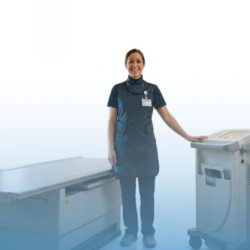
A mammography unit is a rectangular box housing the x-ray tube. We use this dedicated equipment only for breast imaging; its accessories expose just the breast to x-rays. An attached device holds and compresses the breast and positions it for imaging at different angles.
How the Exam Is Performed
A qualified radiology technologist positions you and places the breast on a platform, then compresses it with a paddle (often clear Plexiglas or similar plastic). Compression:
- Evens out breast thickness so radiologists can visualize all the tissue;
- Spreads out tissue so overlying breast tissue doesn’t hide small abnormalities;
- Allows a lower x-ray dose, since the machine images a thinner amount of tissue;
- Holds the breast still to prevent motion blur;
- Reduces x-ray scatter for a sharper image.
The technologist steps behind a glass shield to make the exposure, sending a beam of x-rays through the breast to the film or detector behind the plate. You’ll change position slightly between images — typically a top-to-bottom view and a side view — and the process repeats for the other breast.
The whole exam takes about half an hour. Afterward, you’ll wait briefly while the technologist checks whether they need any additional images.
Who Interprets the Results, and How Do I Get Them?
A radiologist — a physician experienced in mammography and other x-ray exams — reviews the images, describes any abnormalities, and suggests a likely diagnosis. The radiologist dictates a report and sends it to your referring physician, who will share your results with you.
Also Read: Pediatric abdominal ultrasound and preparation
Limitations of Mammography
Interpreting mammograms can be difficult, since a normal breast looks different from woman to woman. Powder or salve on the breast, or a history of breast surgery, can also affect how the image appears. Because some breast cancers are hard to visualize, a radiologist may compare the current image against previous exams — mammography can’t show every breast cancer.
Breast implants can also make readings harder. Both silicone and saline implants are opaque on x-rays, and they can block a clear view of the tissue behind them, especially when the implant sits in front of rather than beneath the chest muscle. Patients with implants should let the technologist know beforehand.
Source: Canadian Association of Radiologists Patient Information
Ready to book your mammogram? Visit our Mammography & OBSP service page for eligibility and booking details, or book an appointment directly.
Related reading: When Should You Get Your First Mammogram in Ontario? OBSP Age & Eligibility Guidelines | What Is OBSP? A Complete Guide to the Ontario Breast Screening Program at Annex Medical Imaging | Breast cancer screening guidelines.
Related Reading
continue reading
Related Posts
How long do X-ray and ultrasound results take in Toronto? Learn who reads your images, when your doctor gets the report, and what to do while you wait.
Sprain or fracture? Learn when an ankle, wrist, hand or foot X-ray is needed, when to go to urgent care, and how to get a walk-in X-ray in Toronto.

How much radiation is in an X-ray? Plain-language answers on dose, safety and pregnancy from a walk-in X-ray clinic in Toronto.