
At a Glance
- Uses sound waves to measure blood flow speed and direction — no radiation, no needles
- Common for suspected blood clots (DVT), carotid narrowing, varicose veins, and PAD
- Appointment typically takes 20–45 minutes
- A requisition is generally required for OHIP coverage
- Results usually reach your doctor within 24–48 hours (urgent findings same day)
If your doctor has ordered a vascular Doppler ultrasound — sometimes called a “vascular ultrasound” or “Doppler study” — you might be picturing something more complicated or intimidating than it actually is. In reality, it’s a painless, non-invasive scan that uses sound waves to look at how blood is moving through your arteries and veins — no needles, no radiation, no dye. It’s also one of the most useful tools available for catching circulation problems before they progress into something more serious, like a blood clot or a stroke.
This guide walks through what a vascular ultrasound actually measures, the symptoms that typically lead to a referral, how to prepare depending on which vessels are being studied, and what the appointment itself feels like.
What a Vascular Doppler Ultrasound Actually Checks
Unlike a general ultrasound that mostly looks at organ structure, a vascular ultrasound (also called a Doppler ultrasound) measures the speed and direction of blood flow using the Doppler effect — the same principle used in radar to measure the speed of a moving object. It’s commonly used to investigate:
- Deep vein thrombosis (DVT) — blood clots in the leg veins, which can be dangerous if a piece breaks off and travels to the lungs
- Carotid artery narrowing — a buildup of plaque in the neck arteries that’s a significant risk factor for stroke
- Varicose veins and venous insufficiency — when the valves in your leg veins aren’t working properly, allowing blood to pool
- Peripheral arterial disease (PAD) — reduced blood flow to the arms or legs, often due to atherosclerosis
- Follow-up monitoring after vascular surgery, stenting, or a dialysis fistula, to confirm everything is functioning as expected
Common symptoms that lead to a referral
Doctors typically order a vascular ultrasound in response to specific symptoms or exam findings, including leg swelling (especially if it’s one-sided), calf pain or tenderness, skin discoloration or warmth over a vein, cramping or aching when walking that resolves with rest (a classic sign of PAD called claudication), dizziness or a prior mini-stroke (TIA), or a whooshing sound (called a bruit) that your doctor hears with a stethoscope over an artery.
How to prepare, by study type
Preparation depends heavily on which vessels are being studied, so it’s worth understanding the differences:
- Leg (venous or arterial) studies: No special preparation is needed. Wear loose, comfortable clothing that can be easily rolled up or removed around the legs.
- Carotid (neck) studies: No fasting is required, and you can eat and take medications normally beforehand.
- Abdominal vascular studies (e.g., renal artery or aorta): Your technologist may ask you to fast for several hours beforehand, since bowel gas can interfere significantly with image quality in the abdomen.
Because prep instructions can vary by the exact study your doctor ordered, it’s always worth confirming specific instructions with our front desk when you book or check in.
What to expect during the scan
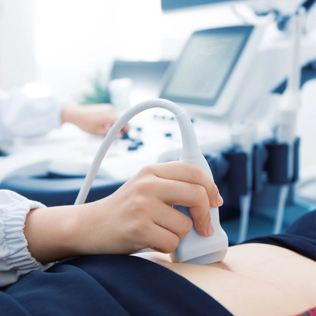
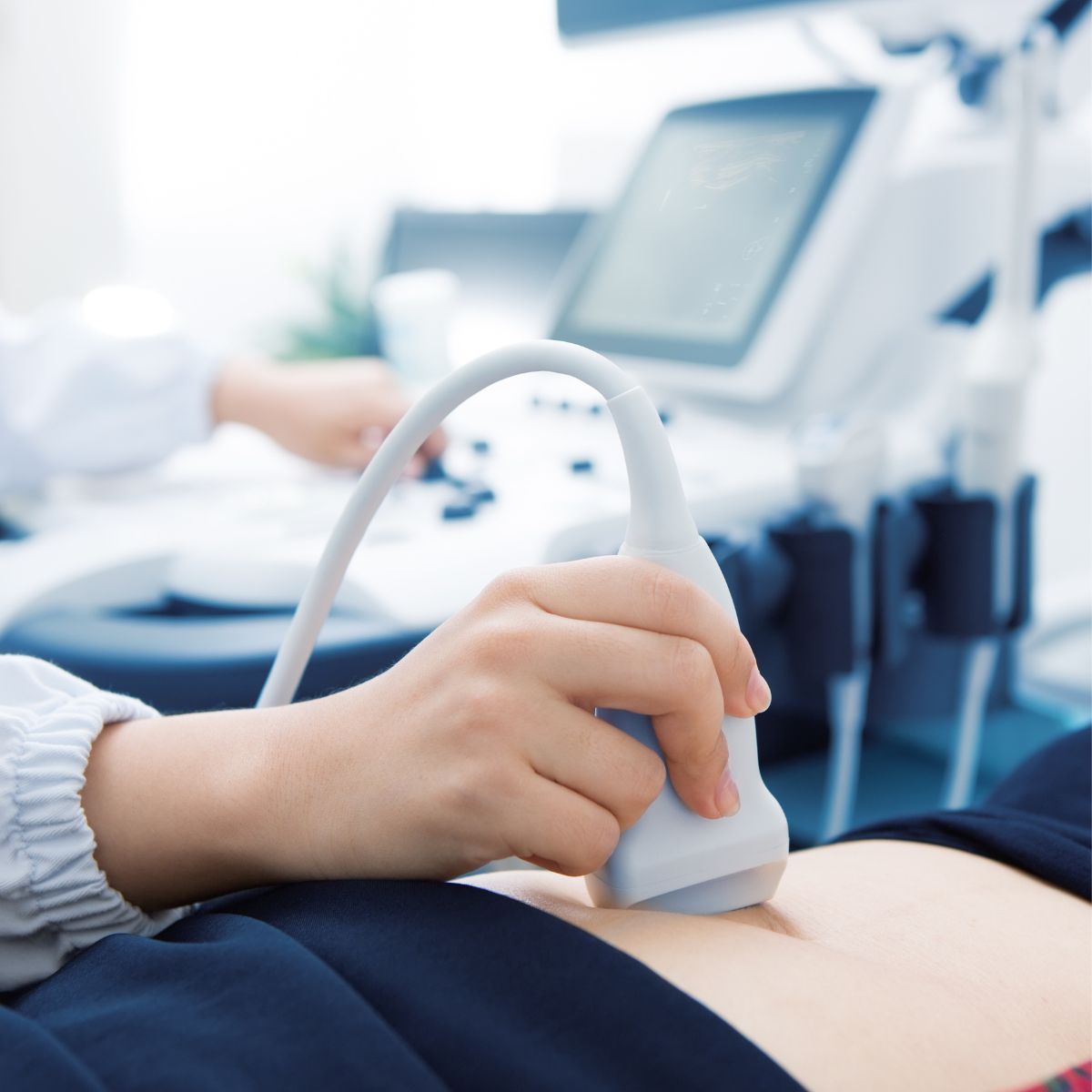
A certified sonographer applies a small amount of gel to the area being studied and moves a handheld probe (called a transducer) across your skin. You may hear a pulsing “whoosh” sound through the machine’s speakers — that’s the sound of your own blood flow being converted into audible feedback, which helps the sonographer assess flow patterns in real time. The exam is entirely painless, though the sonographer may need to press somewhat firmly in certain spots to get a clear reading. Depending on how many vessels are examined, the appointment typically takes 20–45 minutes.
What happens if something abnormal is found
If the sonographer’s images and the interpreting radiologist’s read suggest a blood clot, significant narrowing, or another concern, your referring physician is notified promptly, and urgent findings are typically communicated the same day given the potential seriousness of conditions like DVT. Your doctor will then discuss next steps with you directly, which might include medication, further imaging, or a referral to a vascular specialist.
How vascular ultrasound compares to other vascular imaging tests
Vascular ultrasound is usually the first-line test for suspected circulation problems because it’s fast, painless, involves no radiation or contrast dye, and can be performed on a walk-in or near-walk-in basis. By comparison, tests like CT angiography or MRI angiography involve either radiation or a longer scheduling process, and often require an injected contrast dye that isn’t suitable for everyone (for example, patients with certain kidney conditions or contrast allergies). For most day-to-day concerns — a swollen calf, a suspected DVT, carotid narrowing, or leg pain with walking — ultrasound gives your doctor enough information to make a treatment decision without needing a more invasive or resource-intensive test. Your doctor may still order a CT or MRI angiogram afterward in more complex cases, but ultrasound is almost always the sensible starting point.
Vascular disease often has no symptoms until it’s advanced — which is exactly why imaging plays such an important role.
Related Reading
- General Abdominal Ultrasound: What It Checks and How to Prepare (Fasting Guide)
- Obstetric Ultrasound in Toronto: What Happens at Your First, Anatomy & Growth Scans
Frequently asked questions
Is a vascular ultrasound painful?
No — it’s a non-invasive, painless test. Some patients feel mild pressure from the probe, but nothing more.
How long does a vascular ultrasound take?
Typically 20–45 minutes, depending on how many vessels or areas need to be examined.
Do I need a referral for a vascular ultrasound?
Yes, a requisition from a physician or nurse practitioner is generally required for OHIP coverage.
Can I drive myself home afterward?
Yes — there’s no sedation or contrast involved, so you can resume normal activities immediately.
What’s the difference between a vascular ultrasound and a regular ultrasound?
A regular (general) ultrasound looks primarily at organ structure, while a vascular ultrasound specifically measures blood flow speed and direction using Doppler technology.
Will I need to remove compression stockings before the scan?
Yes, if you wear compression stockings, plan to remove them before your appointment so the sonographer has clear access to the area being examined.
Can a vascular ultrasound detect a blood clot that’s already travelled to the lungs?
No — a leg or arm vascular ultrasound checks the vessels being scanned directly; a suspected clot in the lungs (pulmonary embolism) requires different imaging, typically a CT scan, usually arranged urgently through a hospital.
Additional resources on vascular health
- Heart & Stroke Foundation of Canada — Information on stroke, heart disease, and vascular risk factors
- Thrombosis Canada – Patient & Family Information — Patient resources on blood clots (DVT/PE) and vascular disease
- Ontario – Wait Times for Diagnostic Imaging — Check average wait times for vascular ultrasound and other imaging in Ontario
Why it matters
Vascular disease often has no symptoms until it’s advanced, which is exactly why imaging plays such an important role — catching narrowed arteries or a developing clot early can mean the difference between a simple, manageable treatment plan and a medical emergency. If your doctor has ordered a vascular ultrasound, Annex Medical Imaging offers general, obstetric, and vascular ultrasound with certified sonographers at our Bathurst Street location, with most appointments available within 1–2 business days.
continue reading
Related Posts

What is OBSP? A complete guide to Ontario's free breast cancer screening program — eligibility, self-referral, what to expect, and how results work.

Wondering when you should start getting mammograms in Ontario? Here's what changed with OBSP eligibility and when screening typically begins.

Find Annex Medical Imaging at 800 Bathurst St, a 1-minute walk from Bathurst Station. Directions, parking, and hours for your Toronto imaging appointment.