
At a Glance
- X-rays show bone alignment clearly but not soft tissue like ligaments or tendons
- Knee X-rays are often taken standing (weight-bearing) for a more accurate view
- Hip X-rays after a fall in older adults are a same-day priority
- Multiple views are usually taken for a complete picture
- No appointment needed — walk in with or without a requisition
Joint pain is one of the most common reasons people end up in a walk-in X-ray clinic. Whether it’s a sports injury, a fall, or pain that’s crept up gradually over months, an X-ray is often the first imaging test your doctor reaches for, because it’s fast, widely available, and excellent at showing bone alignment and structure. Here’s a closer look at why shoulder, knee, and hip X-rays are so commonly ordered — and what actually happens once you’re in the room.
Shoulder X-rays
Shoulder X-rays are commonly ordered for suspected fractures after a fall onto an outstretched arm, dislocations (which are relatively common at the shoulder given its wide range of motion), persistent shoulder pain that hasn’t responded to rest, or as part of an arthritis assessment in older adults. An X-ray shows bone alignment clearly and can rule out fracture or dislocation quickly, but it’s important to know that soft-tissue issues like rotator cuff tears, labral tears, or tendinitis won’t show up directly on an X-ray — those typically require ultrasound or MRI as a follow-up if the X-ray comes back normal but symptoms persist.
Knee X-rays
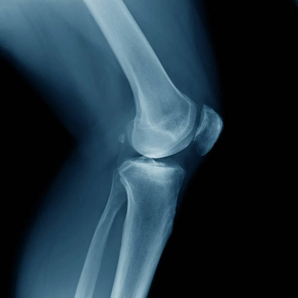
Knee X-rays are typically ordered after twisting injuries, direct trauma (like a fall or a collision during sport), persistent swelling, or to assess arthritis and joint space narrowing in patients with chronic knee pain. Interestingly, knee X-rays are often taken standing up (called weight-bearing views) rather than lying down, because this gives a much more accurate picture of how the joint behaves and how much cartilage space remains under normal, everyday load — a knee X-ray taken lying down can sometimes look deceptively normal compared to one taken standing.
Hip X-rays
A hip X-ray is often the very first step after a fall in older adults, specifically to rule out a hip fracture, which is one of the more serious injuries in this age group and requires urgent evaluation. Hip X-rays are also used to investigate hip or groin pain in younger, active patients, and to evaluate arthritis and joint degeneration over time. Because hip fractures are a genuine medical priority requiring rapid decision-making, walk-in access to X-ray matters enormously here — getting imaged quickly can meaningfully speed up treatment decisions and reduce complications from delayed diagnosis.
What to Expect During Your Shoulder, Knee, or Hip X-Ray
- You may be asked to change into a gown or remove jewellery and clothing with zippers or buttons near the joint being imaged
- Multiple views (typically front, side, and sometimes an angled view) are taken for a complete picture, since a single view can hide certain fractures or abnormalities
- The scan itself takes just a few minutes; positioning — especially for knee and shoulder views — can take a bit longer to get right
- Radiation exposure is low, and our technologists use precise, targeted imaging with shielding where appropriate
- If you’re in significant pain, let the technologist know beforehand so they can help position you as comfortably as possible
What happens if a fracture is found
If the radiologist identifies a fracture or another significant finding, this is typically flagged urgently to your referring provider so you can be directed to appropriate care quickly — whether that’s a cast, a splint, or a referral to orthopedics or the emergency department, depending on severity. Annex Medical Imaging doesn’t provide treatment directly, but fast, accurate imaging is often the critical first step in getting you the right care fast.
Other joints we commonly image
While shoulder, knee, and hip X-rays are among the most frequent walk-in requests, the same general principles apply to other joints too. Ankle and foot X-rays are common after twisting injuries or falls, particularly in athletes and active kids. Wrist and hand X-rays are frequently ordered after a fall onto an outstretched hand, a common mechanism for fractures like a scaphoid or distal radius fracture. Elbow X-rays are less common but are used to investigate suspected fractures after falls or persistent pain from repetitive strain. In every case, the same walk-in process applies — arrive, check in, and be seen by a certified technologist.
Arthritis staging and monitoring over time
For patients with diagnosed or suspected arthritis, X-rays aren’t just a one-time diagnostic tool — they’re often repeated periodically to monitor how joint space and bone changes progress over months or years. Comparing a new X-ray to previous images can help your doctor or a specialist assess whether a treatment plan is working or whether the joint is deteriorating faster than expected, which can influence decisions about medication, physiotherapy, or eventually surgical options like joint replacement.
Because hip fractures are a genuine medical priority requiring rapid decision-making, walk-in access to X-ray matters enormously here.
Related Reading
- DEXA Scan vs. Bone Mineral Density (BMD) Test: Are They the Same Thing?
- Ultrasound vs. X-Ray vs. Mammogram: Which Imaging Test Do You Actually Need?
Frequently asked questions
Do I need a referral for a joint X-ray?
In many cases no, though having one from your doctor helps ensure your results are directed to the right place for follow-up care.
Will an X-ray show a torn ligament or tendon?
No — X-rays show bone, not soft tissue. Ligament and tendon injuries typically require ultrasound or MRI.
Can I walk in for an X-ray the same day as a sports injury?
Yes, same-day walk-in access is one of the main advantages of a dedicated imaging clinic over waiting for a scheduled hospital appointment.
Why does my knee X-ray need to be taken standing up?
Standing (weight-bearing) X-rays show how the joint behaves under normal load, giving a more accurate picture of cartilage space and alignment than a scan taken lying down.
Is radiation exposure from a joint X-ray a concern?
The radiation dose from a routine joint X-ray is very low, and our technologists use precise, targeted techniques to minimize exposure further.
Additional resources on joint health and imaging
For more on diagnostic imaging access and joint health in Ontario and Canada, these resources go into further detail:
- Wait Times for Diagnostic Imaging — Government of Ontario’s official wait time reporting for X-ray and other imaging
- Choosing Wisely Canada – Orthopaedics — evidence-based guidance on when imaging is and isn’t needed for joint injuries
- Bone & Joint Canada — patient education on osteoarthritis and joint health
No appointment necessary
Most X-ray exams at Annex Medical Imaging don’t require a booked appointment — walk in with your requisition (or without one, in many cases) and a certified technologist will see you. Results are typically with your referring provider within 24–48 hours.
continue reading
Related Posts

What is OBSP? A complete guide to Ontario's free breast cancer screening program — eligibility, self-referral, what to expect, and how results work.

Wondering when you should start getting mammograms in Ontario? Here's what changed with OBSP eligibility and when screening typically begins.

Find Annex Medical Imaging at 800 Bathurst St, a 1-minute walk from Bathurst Station. Directions, parking, and hours for your Toronto imaging appointment.