")
News, Articles and Information
Preparation and updates
Explore the procedure and preparation of each test as well our blogs and news
pelvic ultrasound is most often used to examine the uterus and ovaries and, during pregnancy, to monitor the health and development of the embryo or fetus
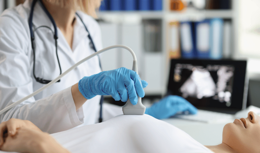
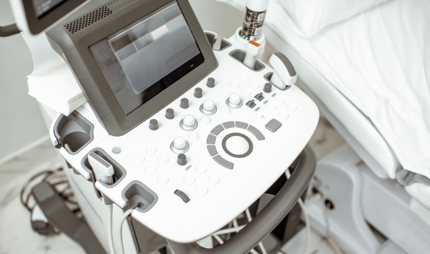
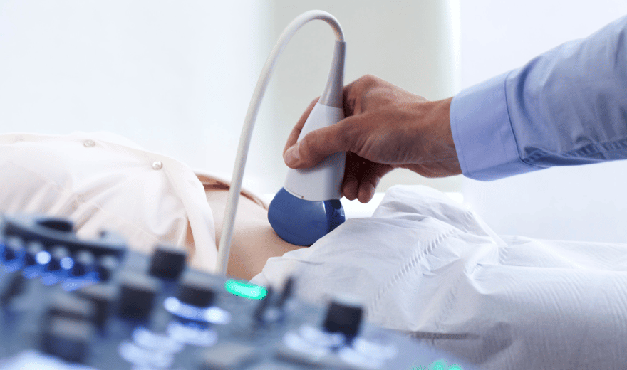
Ultrasound imaging, also called ultrasound scanning or sonography, involves exposing part of the body to high-frequency sound waves to produce pictures of the inside of the body.
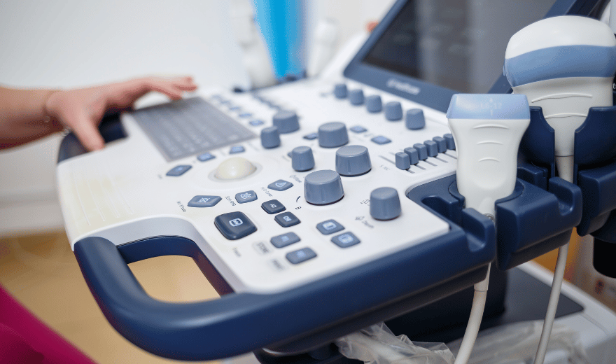
Ultrasound or sonography involves the sending of sound waves through the body. Those sound waves are reflected off the internal organs.
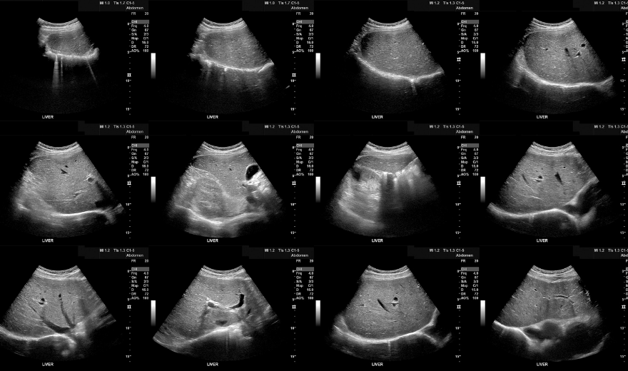
An abdominal ultrasound image is a useful way of examining internal organs, including the liver, gallbladder, spleen, pancreas, kidneys, and bladder.
If an ultrasound is required early in your pregnancy, you will be required to have a full bladder for the procedure.
-
In cases when the appendix is not clearly visualized during a clinic-based ultrasound, a hospital-based ultrasound as an outpatient may be needed.

Ultrasound imaging of the scrotum is the primary imaging method used to evaluate disorders of the testicles and surrounding areas.

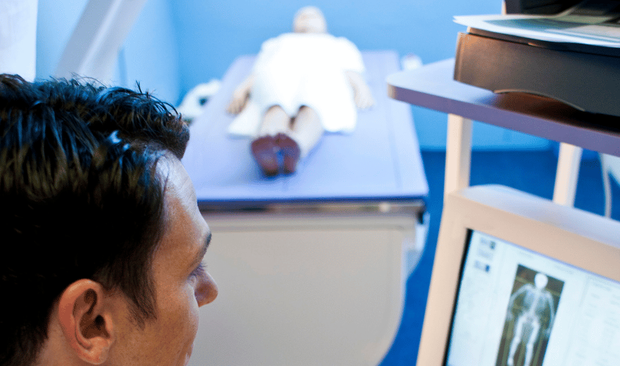
Cancer screening is testing done on people who may be at risk of getting cancer, but who have no symptoms and generally feel fine.