
WHAT IS RADIOGRAPHY-BASED (X-RAY) BONE MINERAL DENSITOMETRY (BMD)?
Every day, radiologists (physicians specially trained to diagnose conditions and diseases by obtaining and interpreting medical images) use radiography, or x-rays, to view and evaluate bone fractures and other injuries of the musculoskeletal system. However, a plain x-ray test is not the best way to assess bone density. To detect osteoporosis accurately, radiologists use an enhanced form of x-ray technology called dual-energy x-ray absorptiometry (DXA or DEXA). DEXA bone densitometry is today’s established standard for measuring bone mineral density (BMD). DEXA is a quick, painless procedure for measuring bone loss. Measurement of the lower spine and hips is most often done.
WHAT ARE SOME COMMON USES OF THE PROCEDURE?
Bone Mineral densitometry (BMD) is used most often to diagnose osteoporosis, a condition that often affects women after menopause, but may also be found in men. Osteoporosis involves a gradual loss of calcium and structural changes, causing the bones to become thinner, more fragile, and more likely to break. The BMD test can also assess your risk for developing fractures. If your bone density is found to be low, you and your physician can work together on a treatment plan to help prevent fractures before they occur. BMD testing is also effective in tracking the effects of treatment for osteoporosis or for other conditions that cause bone loss.
HOW SHOULD I PREPARE FOR THE PROCEDURE?
On the day of the exam, eat normally, but don’t take calcium supplements for at least 24 hours beforehand. Please wear loose, comfortable clothing, avoiding garments that have zippers, belts, or buttons made of metal. Inform your physician and staff at Annex Medical Imaging if you recently had a barium examination or have been injected with a contrast material for a computed tomography (CT) scan or radioisotope scan; you may have to wait 10-14 days before undergoing a DEXA test. Women should always inform their physician, radiologist, and x-ray technologist if there is a possibility they are pregnant.
HOW IS THE PROCEDURE PERFORMED?
BMD testing takes about 20 minutes. You may be asked to undress and put on a hospital gown. Then you will lie on a padded table with an x-ray generator below and a detector (an imaging device) above.
Most often radiologists focus on bone loss in the spine and hip where most osteoporosis-related fractures happen. During an examination of the spine, your legs will be supported on a padded box to flatten your pelvis and lower (lumbar) spine. To assess your hip, the technologist will place your foot in a device that rotates the hip inward. In both cases, the detector is slowly passed over the area, generating images on a computer monitor.
WHO INTERPRETS THE RESULTS AND HOW DO I GET THEM?
The results of the exam are interpreted by a radiologist (a physician specially trained to diagnose conditions and diseases by obtaining and interpreting medical images). The radiologist will send an interpretation of your results and a signed report to your primary care physician who will work with you to develop a treatment plan.
WHAT DOES THE BMD EQUIPMENT LOOK LIKE?
The BMD unit measures bone density in the hip and spine. The BMD unit has a large, flat table and an “arm” suspended overhead. During the x-ray exposure the arm moves over the patient’s body. How does the procedure work?
The BMD unit sends a thin beam of low-dose x-rays through the area of the body being examined. The radiation is absorbed by a detector and information is sent to a computer.
All BMD units feature special software to compute the data and display them on a computer monitor, allowing your radiologist to make an accurate diagnosis. The amount of radiation used is extremely small—less than one tenth the dose of a standard chest x-ray.
WHAT WILL I EXPERIENCE DURING THE X-RAY PROCEDURE?
BMD testing is a simple, non-invasive procedure. Once on the table, you may be asked to hold an awkward position for a short time while the arm of the machine passes over your body taking measurements. It is important that you stay as still as possible during the procedure to ensure a clear, useful image. No anesthesia is required. The procedure is painless, and radiation exposure is minimal.
WHAT ARE THE BENEFITS VS. RISKS?
Benefits
MD test is the most accurate method available for the diagnosis of osteoporosis. It is also considered an accurate estimator of fracture risk. It will not tell whether you will or will not have a fracture, but gives relative risk of suffering a fracture, just as cholesterol and blood pressure help determine risk for heart disease. A low reading should not cause you to be anxious, but may help you set healthy goals. As with other diseases and conditions, early detection is the key to prevention of further bone loss and eventual fractures.
Risks
No complications are expected with BMD testing.
WHAT ARE THE LIMITATIONS OF BMD TESTING?
Despite its effectiveness as a method of measuring bone density, it is of limited use in people with a spinal deformity or those who have had previous spinal surgery. The presence of vertebral compression fractures or osteoarthritis may interfere with the accuracy of the test. CT scans may be more useful in such instances.
Source: Canadian Association of Radiologists Patient Information
Preparation
BONE MINERAL DENSITOMETRY
Bone Mineral Density (BMD) is a test which detects low bone density. The most common bone density test is called dual energy x-ray absorptiometry (DEXA).
Dual-energy x-ray absorptiometry (DEXA) is an imaging technology that uses a very low amount of x-ray energy to detect the presence of osteoporosis. Osteoporosis is a disease that gradually weakens bones, leading to bone fragility and an increased chance of fractures to the spine, hips and wrists. This weakening may be due to aging or caused by other risk factors that combine with age. Often called the “silent disease,” osteoporosis rarely shows symptoms until excessive bone mass has been lost. The most visible symptoms may include loss of height along with curvature of the upper back. DEXA scanning can identify low bone density in patients at an early stage, enabling doctors to prescribe appropriate treatment before the condition worsens. Images of the lower spine and hips are most often used in checking for osteoporosis.
BONE MINERAL DENSITOMETRY PREPARATION
These instructions are IMPORTANT. Please follow them.
- On the day of examination DO NOT take calcium supplements or iron tablets until after the examination is completed. These supplements may be found in multi-vitamins and antacids. Please be certain to read the labels when taking all medications within 24 hours prior to the examination. Dairy products are acceptable.
- You should not have certain radiologic studies within the 7 days before the exam, i.e., barium studies, studies involving IV or oral contrast, or Nuclear Medicine studies. That waiting period is important to prevent any residual barium from interfering with your DEXA exam.
- It is preferable to wear clothing without zippers or fasteners (e.g. jogging suit or leggings).
- Please let us know before your exam begins if you may be pregnant.
continue reading
Related Posts
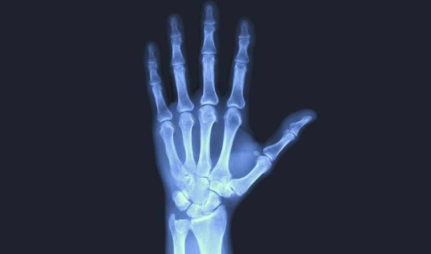
Radiography equipment consists of a large, flat table with a drawer that holds a tray into which an x-ray film cassette is placed.
involves sending sound waves into the body. These sound waves reflect off the internal organs and are recorded by special instruments that create images of anatomic parts.
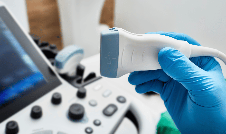
A radiologist (a physician experienced in ultrasound and other radiology examinations) will analyze the images and send a signed report with his or her interpretation to the patient’s personal physician.