
WHAT IS VASCULAR ULTRASOUND IMAGING?
Ultrasound or sonography involves the sending of sound waves through the body. Those sound waves are reflected off the internal organs. The reflections are then interpreted by special instruments that subsequently create an image of anatomic parts. No ionizing radiation (x-ray) is involved in vascular ultrasound imaging.
An ultrasound image is a useful way of evaluating the body’s circulatory system. Ultrasound images are captured in real-time, so they can help radiologists monitor the blood flow to organs and tissues throughout the body, as well as evaluate the placement and success of repair, such as after arterial bypass surgery. With ultrasound images, radiologists can locate and identify blockages (stenosis) and abnormalities like blood clots, plaque or emboli, and help plan for their effective treatment.
WHAT ARE SOME COMMON USES OF THE PROCEDURE?
Ultrasound imaging of the body’s veins and arteries can help the radiologist see and evaluate blockages to blood flow, such as clots in veins and plaque in arteries. With knowledge about the arterial blood flow gained from an ultrasound image, the radiologist can often determine whether a patient is a good candidate for a procedure like angioplasty. Ultrasound images may also be used to plan or review the success of procedures that graft or bypass blood vessels—such as renal (relating to the kidney) artery bypass. Ultrasound of the veins may reveal blood clots that require treatment such as anticoagulant therapy (blood thinner), or filters to prevent clots from traveling to the lungs (embolism).
HOW SHOULD I PREPARE FOR THE PROCEDURE?
You should wear comfortable, loose-fitting clothing for your ultrasound exam. No other preparation is required. If your abdominal vessels are being studied, you will need to fast before the procedure.
HOW DOES THE PROCEDURE WORK?
Ultrasound imaging is based on the same principles involved in the sonar used by bats, ships at sea, and anglers with fish detectors. As a controlled sound bounces against objects, its echoing waves can be used to identify how far away the object is, how large it is, its shape and its internal consistency (fluid, solid or mixed).
The ultrasound transducer functions as both a loudspeaker (to create the sounds) and a microphone (to record them). When the transducer is pressed against the skin, it directs a stream of inaudible, high-frequency sound waves into the body. As the sound waves echo from the body’s fluids and tissues, the sensitive microphone in the transducer records tiny changes in the sound’s pitch and direction. These signature waves are instantly measured and displayed by a computer, which in turn creates a real-time picture on the monitor. Still frames of the moving picture are “frozen” to capture a series of images. Blood flow changes the pitch of the sound beam; this Doppler effect can be heard or detected on the image as color or displayed graphically.
WHAT WILL I EXPERIENCE DURING THE PROCEDURE?
Most ultrasound studies are fast and easy. You will lie on your back on an examining table. The sonographer or radiologist will spread some gel on your skin and then press the transducer firmly against your body, moving it until the desired images are captured. Most exams take less than 30 minutes; however, more complicated examinations may take somewhat longer.
WHAT ARE THE BENEFITS VS. RISKS?
Benefits
- Ultrasound scanning is non-invasive (no needles or injections) and usually painless.
- Ultrasound is widely available and easy to use.
- Ultrasound uses no ionizing radiation.
- Ultrasound images can demonstrate structure, movement and function in the body’s blood vessels in real time.
Risks
- For standard diagnostic ultrasound there are no known harmful effects on humans.
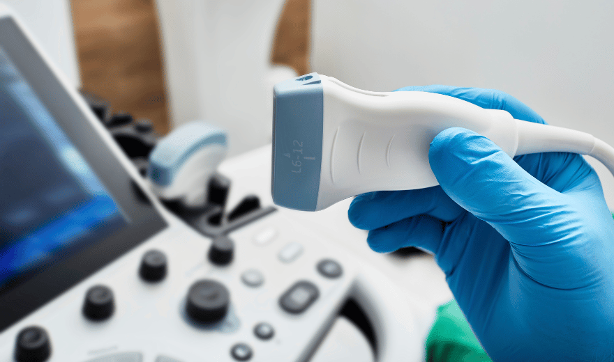
WHAT DOES THE EQUIPMENT LOOK LIKE?
The equipment consists of a transducer and a monitoring system. The transducer is a small hand-held device that resembles a microphone. The radiologist or sonographer spreads a lubricating gel on the area being examined, and then presses this device firmly against the skin.
The transducer passes the image to the ultrasound machine and from there to a viewing monitor. The radiologist or ultrasonographer watches this screen during an examination and captures representative images for storage. Often, the patient is able to see it as well. Blood flow also produces sound that can be heard with Doppler ultrasound. You may also hear the sounds.
HOW IS THE PROCEDURE PERFORMED?
The patient is positioned on an examination table and a clear gel is applied to the area that will be examined. The gel helps the transducer make a secure contact and eliminates air pockets between the transducer and the skin, since the sound waves cannot penetrate air. The sonographer, vascular technologist or radiologist then presses the transducer firmly against the skin and sweeps along the area of interest, reviewing the images on the monitor and capturing “snapshots” as required. For venous ultrasound examinations, the transducer is pressed gently on the leg or arm.
When the examination is complete, the patient may be asked to dress and wait while the ultrasound images are reviewed, either on film or on a monitor. Often, though, the sonographer or radiologist is able to review the ultrasound images in real time as they are acquired, and the patient can be released immediately.
WHO INTERPRETS THE RESULTS AND HOW DO I GET THEM?
A radiologist (a physician experienced in ultrasound and other radiology examinations) will analyze the images and send a report to your personal physician. Generally, you will receive results of the examination from your referring physician
WHAT ARE THE LIMITATIONS OF VASCULAR ULTRASOUND IMAGING?
- Vessels deep in the body are harder to see than superficial vessels. Specialized equipment may be necessary.
- Calcifications that occur as a result of atherosclerosis may obstruct the ultrasound beam.
- Sometimes the ultrasound cannot tell the difference between a blood vessel that is closed or very nearly closed because the weak volume of blood flow produces a weak signal.
Preparation
VASCULAR ULTRASOUND
Ultrasound or sonography involves the sending of sound waves through the body. Those sound waves are reflected off the internal organs. The reflections are then interpreted by special instruments that subsequently create an image of anatomic parts. No ionizing radiation (x-ray) is involved in ultrasound imaging.
An ultrasound image is a useful way of evaluating the body’s circulatory system. Ultrasound images are captured in real-time, so they can help radiologists monitor the blood flow to organs and tissues throughout the body, as well as evaluate the placement and success of repair, such as after arterial bypass surgery. With ultrasound images, radiologists can locate and identify blockages (stenosis) and abnormalities like blood clots, plaque or emboli, and help plan for their effective treatment.
VASCULAR ULTRASOUND PREPARATION
These instructions are IMPORTANT. Please follow them.
- You should wear comfortable, loose-fitting clothing for your ultrasound exam. No other preparation is required.
- If your abdominal vessels are being studied, you will need to fast before the procedure.
continue reading
Related Posts
A radiologist (a physician experienced in ultrasound and other radiology examinations) will analyze the images and send a signed report with his or her interpretation to the patient’s personal physician.